As part of the AIMIRA project, the University of Groningen and the University Medical Center Groningen are developing a patient-specific "digital twin" of the rectal area, designed to assist surgeons during robot-assisted minimally invasive procedures.
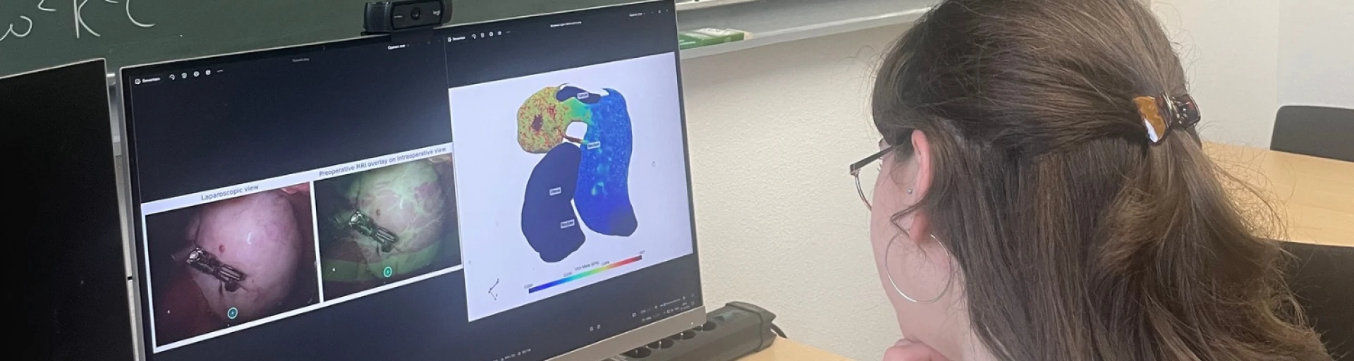
During surgery, the positioning of organs differs from that seen on preoperative MRI scans—due to factors such as the patient’s orientation, the insufflation of gas, and the patient’s breathing. Consequently, the 3D model must provide a real-time visualization of the tumor’s location and the position of vulnerable nerves.
The model relies primarily on conventional computational methods, employing artificial intelligence only for patient characteristics that are difficult to measure directly; this ensures that the results remain transparent and their derivation can be clearly explained.
A key milestone has already been reached: the model can now account for the effects of gravity on organ movement. The next phase involves integrating the model with surgical instruments.
The project is expected to run until 2030, with financial backing from partners including Intuitive. Following this, the technology will undergo clinical testing and certification before it can be implemented for use in the operating room.
AIMIRA can help surgeons operate with greater precision using a digital twin of the patient
Surgeons can benefit enormously from new computer technologies, enabling them to work more accurately and efficiently during minimally invasive surgery – commonly known as keyhole surgery. In the AIMIRA project, the University of Groningen (RUG) and the University Medical Centre Groningen (UMCG) are jointly developing a personalised digital twin of the area around the rectum. In the long term, this application could help a surgeon to operate more accurately and quickly in a dynamic environment with fewer operating theatre staff. This will enable better and more efficient care to be provided.
The AIMIRA project receives support from Holland High Tech under the PPP Innovation Scheme (PPS-I) of the Ministry of Economic Affairs and Climate. This article is the fifth in a series of articles on the impact of innovation through PPS-I projects.
* Photo credit: Rijksuniversiteit Groningen
Surgery in the lower pelvic region is extremely complex. There is very little room to manoeuvre, and important nerves are present in the area. If these are damaged, it can lead to serious complications, such as incontinence and sexual dysfunction. In current practice, the surgeon uses the patient’s MRI scans to prepare for the operation. The surgeon then performs the procedure using real-time video images transmitted by the surgical instruments. However, above all, a great deal of knowledge and experience is required. One challenge with imaging is that the patient lies flat during the MRI scans but is tilted slightly to one side during the operation. Furthermore, gas is pumped into the abdomen. All of this is done to create maximum manoeuvrability for the surgeon.
As a result, during the operation, the organs in the abdomen have shifted in relation to the MRI images. The surgeon must interpret the information from the MRI scan and apply it to the actual situation during the procedure. To make matters even more complex, the organs and tissues move as a result of the patient’s breathing and the movements of the surgical instruments within the abdomen. “Ultimately, with that puzzle in your head, you have to know where to be vigilant,” explains Esther Consten, Professor of Surgery specialising in gastrointestinal and oncological surgery at the UMCG. Her greatest wish is to address this issue using new technology.
“Ultimately, with that puzzle in your head, you need to know where to be vigilant.”
Esther Consten, Professor of Surgery at the University Medical Centre Groningen
A large image database forms the basis
In 2022, she discussed this idea with Elisabeth Wilhelm, associate professor of Robotics in Healthcare at the University of Groningen. Wilhelm saw the potential to create a patient-specific digital twin of the rectal environment. The AIMIRA project – an acronym for Artificial Intelligence (AI)-driven Minimally Invasive Robot-Assisted Mesorectal Excision – officially launched in the spring of 2025, co-funded by Holland High Tech.
Wilhelm emphasises that preparations for such complex medical projects require a great deal of lead time. Data availability, for example, is a tricky issue. Consten has a very extensive database of medical images which Wilhelm can use to set up the digital twin, but she cannot simply share that information. The project therefore had to comply with the conditions under which the patients had given their consent.
Using the available imaging data, Wilhelm first develops general modelling algorithms. These are then used to create a personalised digital twin for each patient based on pre-operative MRI scans. The aim is for the surgeon to be able to view a 3D image during the operation that highlights the positions of vulnerable tissues and the tumour. This image is updated, partly with the aid of AI, based on real-time images from the camera on the endoscope inserted into the abdomen. “The first milestone has already been reached,” says Wilhelm. “We can now link the effect of gravity to the movement of the abdominal organs within the model.” However, success does depend on many factors, such as the patient’s age, the mobility of the organs – which varies from patient to patient – and the amount of gas present in the abdomen.
“The first milestone has already been reached.”
Elisabeth Wilhelm, Associate Professor of Robotics in Healthcare at the University of Groningen
A digital twin could assist the surgeon with decision-making in real time
Wilhelm explains that, to calculate the mechanics of the tissues, she uses the finite element method. This is a computational method for predicting how complex systems behave under certain conditions. The use of AI is intended to help incorporate patient-specific characteristics that are unknown or difficult to measure. Wilhelm: “However, we try to do this as much as possible using classical models rather than AI. That way, we can explain with a high degree of certainty how the model arrives at its results.”
Once the positioning is working properly, the next step is to link the model to the surgical instruments. “Where are the forces acting on the tissue, and from what angle is the camera viewing it?”
Surgeries are more predictable
Thanks to careful planning, Wilhelm and Consten believe they will be able to make the subsidies from this AIMIRA project last until 2030. Pre-clinical and clinical trials will only be carried out in a follow-up project. Before the technology can be used in everyday operating theatre practice, it must first undergo certification and authorisation processes.
Intuitive, a specialist in robot-assisted surgery, has been working with Esther Consten for many years to achieve the best possible outcomes for all patients, explains Michel Nijs, the company’s regional director for France and the Benelux. Intuitive is therefore providing financial support for this AIMIRA project.
Nijs sees a bright future for this type of development. “Intuitive supports research to contribute to the advancement of scientific and technological knowledge.” More generally, he notes that developments in healthcare technology point to the potential emergence of tools that support real-time decision-making during surgical procedures. Such technology can contribute to greater predictability of procedures and a more efficient use of resources. “In the long term, these developments could lead to an improvement in the quality and efficiency of healthcare. This is a positive development against the backdrop of both staff shortages in the healthcare sector and rising healthcare costs,” says Nijs.
Previous articles in the series:
More news